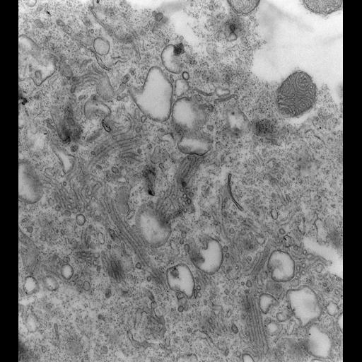
A high resolution image of the changes in the membrane complexes of the Contractile Vacuole Complex. This image illustrates the changes in the CVC caused by cell division and microinjecting mAbs specific for an antigen of the V-ATPase holoenzyme into a P. multimicronucleatum cell. Each condition causes the disappearance of the decorated tubules from the radial arms. In this micrograph the cell was injected with 86 µg/ml of serum containing the DS-1 mAb for the A4 antigen and lightly fixed 45 min later. It is observed that the bundles of decorated tubules had started to round into vesicles, several tubules forming one vesicle. By extrapolation, during bundle formation several decorated tubules apparently arise from each vesicle which accounts for the bundles of tubules emptying into a common duct. The common duct opens into the smooth spongiome. We believe that the V-ATPases have probably lost their functional integrity when the tubules vesiculate as the tubules close to the vesicles in this micrograph no longer have helical decorations. TEM taken on 3/30/93 by M. Ishida with Zeiss 10A operating at 80kV. Neg. 19,800X. Adapted with permission from the J. Cell Sci. 108:693-702, 1993. The raw film was scanned with an Epson Perfection V750 Pro. This image is best used for quantitative analysis. Standard glutaraldehyde fixation followed by osmium tetroxide, dehydrated in alcohol and embedded in an epoxy resin. Microtome sections prepared at approximately 75nm thickness. Additional information available at (http://www5.pbrc.hawaii.edu/allen/).
| Spatial Axis | Image Size | Pixel Size |
|---|---|---|
| X | 4977px | 0.76nm |
| Y | 5558px | 0.76nm |