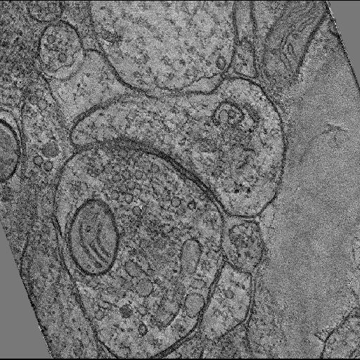
Single computed slice through a tomographic reconstruction of a synaptic contact in hippocampal area CA1 neuropil, from tissue that was prepared by a chemical fixation followed by high pressure freezing.
Full resolution image description
Tar file containing TxBR (projective) combined tomographic volume of high pressure frozen hippocampus tissue, showing a synaptic contact.
Volume_dimension
2242, 3340, 350
Volume scale
0.0008, 0.0008, 0.0008
Animation description
Animation of a tomographic reconstruction of a synaptic contact in hippocampal area CA 1 neuropil, from tissue that was prepared by a chemical fixation followed by high pressure freezing.
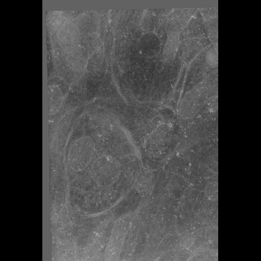
Zero degree tilt electron micrograph from a tilt series of a synaptic contact in hippocampal area CA1 neuropil imaged using intermediate voltage electron microscopy, from tissue that was prepared by a chemical fixation followed by high pressure freezing.
Full resolution image description
Tar file containing original digitized TIFF images, IMOD files (*com, *log, st, preali, fid, rawtlt,...), and TxBR files (*mat, *txt, preali, rawtlt, fid, ...) of high pressure frozen Hippocampus tissue, showing PSD and vesicles.
Animation description
Aligned tilt series of a synaptic contact in hippocampal area CA1 neuropil imaged using intermediate voltage electron microscopy, from tissue that was prepared by a chemical fixation followed by high pressure freezing.