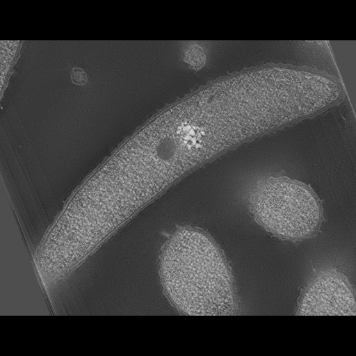
Caulobacter Crescentus specimen from the tilt series shot on a 400kV microscope. This image was taken from the "a" stack at the 0 degree tilt.
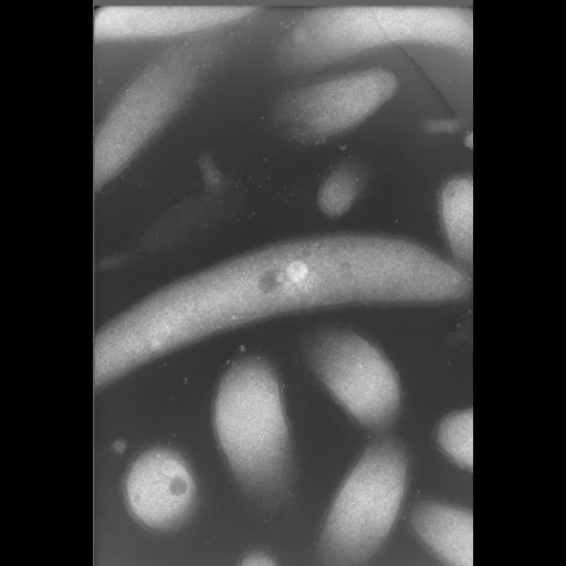
Full resolution image description
a .tar file containing all files needed for reconstruction using the TxBR reconstruction package. Both the a and b stacks, rawtlt file, and TxBR files are included in the .tar file.
Animation description
a .mpg file containing the tilt series. The series shown here is only aligned using cross correlation algorithms.
Produce a series of tomographic data of the normal morphology of Caulobacter Crescentus using rapid freezing methods.
Experimenter(s)
Patrick Viollier
Tom Deerinck
Microscopy product
Microscopy product ID
3660
Instrument
JEOL 4000 #1
Microscopy type
IVEM
Product type
DOUBLE TILT
Image basename
caulodouble2
Spatial Axis
Image Size
Pixel Size
X
2242px
Y
3340px
Subject
Species
Caulobacter
Scientific name
Caulobacter crescentus (NA1000)
Strain
NA1000
Treatment
None
Age class
N/A
Tissue section
Thickness
0.5 µm
Specimen description
Cell type
Caulobacter crescentus
Imaging parameters
Type
Electron microscopy product
Recording medium
film
Magification
30000
Accelerating voltage
400 kV
Specimen preparation
Protocol used
1) Prefix with 1% Acreolin and 2.5% Glutaradehyde2) High Pressure Freezing 3) Free Substitution with 2% Osmium and .1% UA4) Durcupan embedded
Imaging product type
Type
Double tilt
X min range
-60 degrees
X max range
60 degrees
X tilt increment
2 degrees
Y min range
-58 degrees
Y max range
60 degrees
Y tilt increment
2 degrees
Description
Caulobacter Crescentus, 0.5 um thick, 400kv
Citation Information
Lucy Shapiro, Harley McAdams, Patrick H. Viollier, Tom Deerinck, Mason Mackey, John Crum, Mark Ellisman (2003) CCDB:3660, Caulobacter crescentus (NA1000), Caulobacter crescentus. CIL. Dataset. https://doi.org/doi:10.7295/W9CCDB3660