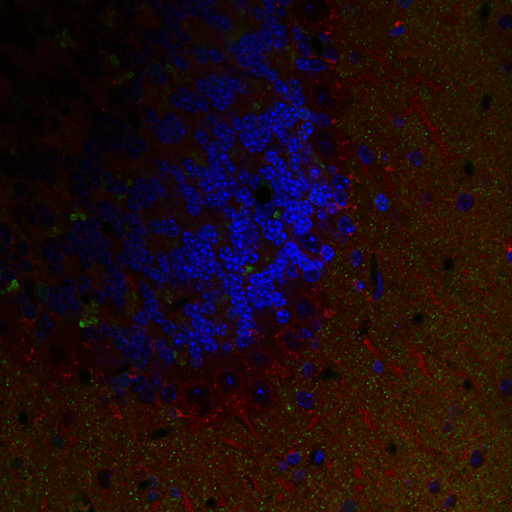
Triple labeled confocal image of the cerebellar cortex of a non-transgenic mouse, immunolabeled for mGluR5 (red), alpha synuclein (green) and counterstained with DAPI (blue) to reveal nuclei. The unevenness in staining likely represents unevenness in the section.
Full resolution image description
Zip archive containing the 3 channel image file in tiff format (112006ccccc_RGB.tiff). Also included is the .oif header file generated by the Olympus Fluoview, which gives additional detail on microscope settings.