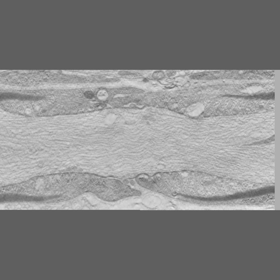
Single slice through a single tilt tomogram of the Node of Ranvier from mouse sciatic nerve prepared by high pressure freezing and freeze substitution of aldehyde fixed material
Full resolution image description
Serial tomographic reconstruction from 3 serial volumes. The resulting volume was mirrored and merged with the original to from a relatively complete node of Ranvier. The downloadable zip file contains the merged volume in Analyze 7.5 format
Volume_dimension
950, 950, 199
Volume scale
0.005, 0.005, 0.008
Animation description
Annotated movie traversing the slices of the serial tomogram and identifying the segmented objects in the surface rendering
Manual segmentation of axonal and glial components of the Node of Ranvier using Xvoxtrace v 2.9. Different parts of the myelin sheath were segmented as different objects. Objects were surfaced using both Synu and Amira
Segmentation file description
Zip archive containing the trace file node_marge2.trace and the segmented objects in synu (*.synu) and Amira (*.iv) format. Also included are several renderings of the surface model. These segmentations were produced from the mirrored volume file to mimic a complete node. That is, the original reconstruction of a sagittal section through a node was mirrored to produce the appearance of a single complete node.