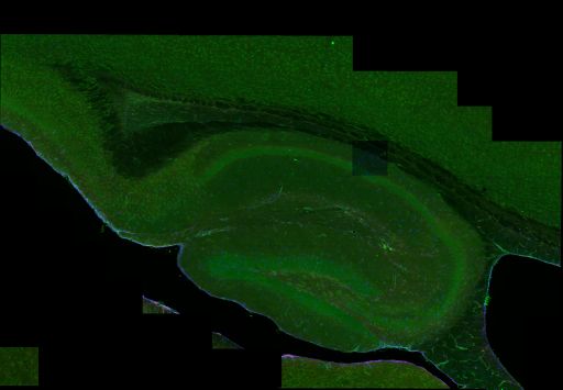
Wide field mosaic of mouse hippocampus immunolabeled for pannexin 1 (green), GFAP (blue) and connexin 43 (red). The section was also counterstained with DAPI (not shown).
Full resolution image description
176 tiles (16x11) were digitally stitched together to generate a 3D mosaic montage of the mouse hippocampus labeled with three different antibodies (Green = Pannexin1 protein, Red = Connexin43 protein, Blue = GFAP protein within astrocytes). This zipped tar file contains 24 reconstructed mosaic images (file names labeled 00-23), one for each of the 24 optical Z-sections that were acquired with a step distance of 0.5 microns. Additionally, the zip file contains a mosaic 2D maximum intensity projection of all 24 Z-sections (file name containing "proj").
Downsampled image description
This image is a mosaic 2D maximum intensity projection of all 24 Z-sections. Voxel scale for this flattened projection of the mosaic hippocampus is 0.41 x 0.41 x 11.5 microns/pixel. The tiles in this image have been normalized, but the colors in the resulting image have not been adjusted.
A neuroinformatics approach to pannexin expression, localization, and interaction with other brain proteins. This research aims to examine brain tissue at high resolution and, in combination with developing neuronformatics tools, to probe for binding partners and signalling partners to investigate the pannexin "interactome". Data submissions for this project will consist of wide-scale high resolution confocal mosaic images of triple-labeled whole mouse brain sections. Pannexin 1 expression will be compared to that of pannexin 2 as well as the connexins. Other putative interaction partners of interest include the P2 purinergic receptors and components of the cytoskeleton.