Alternate header for print version
Advanced search
Contributors
Help
Submit
Search
menu
Cell Process
Cell Component
Cell Type
Organism
Microbial
Alzheimer's
Data Sets
Center for Research in Biological Systems
University of California, San Diego
9500 Gilman Drive
La Jolla, CA 92093-0608, USA
Voice
: (858) 534-0276
Fax
: (858) 534-7497
Email
: dorloff@ncmir.ucsd.edu
Search Results for
David Sabatini
(30 results)
(Not the results you were expecting?)
(Comments)
Still Images
Video/Animation
Z-Stack
Time Series
CIL:41033
NCBI Organism Classification
Rattus rattus
Biological Process
protein sythesis
Cellular Component
ribosome
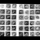
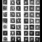
Gallery of transmission electron micrographs of ribosomes purified from a rat liver homogenate by sucrose gradient centrifugation, and negatively stained with uranyl acetate. The 3 columns at left ill...
CIL:41034
NCBI Organism Classification
Rattus rattus
Biological Process
protein sythesis
Cellular Component
polysome
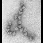
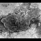
Transmission electron micrograph of a polysome (polyribosome) purified from a rat liver homogenate by sucrose gradient centrifugation, and negatively stained with uranyl acetate. The linear arrangemen...
CIL:41035
NCBI Organism Classification
Rattus rattus
Biological Process
protein sythesis
Cellular Component
polysome
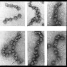
Gallery of transmission electron micrographs of polysomes (polyribosomes) purified from a rat liver homogenate by sucrose gradient centrifugation, and negatively stained with uranyl acetate. The linea...
CIL:41028
NCBI Organism Classification
Rattus rattus
Biological Process
protein sythesis
Cellular Component
ribosome
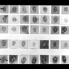
Gallery of transmission electron micrographs of ribosomes purified from a rat liver homogenate by sucrose gradient centrifugation, and negatively stained with uranyl acetate. The gallery illustrates t...
CIL:41029
NCBI Organism Classification
Rattus rattus
Biological Process
protein sythesis
Cellular Component
ribosome
Gallery of transmission electron micrographs of ribosomes purified from a rat liver homogenate by sucrose gradient centrifugation, and negatively stained with uranyl acetate. The 3 columns at right in...
CIL:37200
NCBI Organism Classification
Cavia porcellus
Biological Process
none specified
Cellular Component
mitochondrion
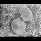
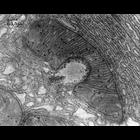
Transmission electron micrograph of guinea pig pancreas mitochondria and lipid droplets. The use of osmium tetroxide as a fixative for electron microscopy was first described by Dr. Palade at the Roc...
CIL:37192
NCBI Organism Classification
Cavia porcellus
Biological Process
none specified
Cellular Component
mitochondrion
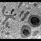
Transmission electron micrograph of mitochondria and lipid droplets from guinea pig pancreas. The use of osmium tetroxide as a fixative for electron microscopy was first described by Dr. Palade at the...
CIL:37193
NCBI Organism Classification
Plasmodium
Biological Process
none specified
Cellular Component
mitochondrion
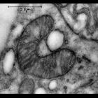
Transmission electron micrograph of a mitochondria from the protist Plasmodium. The use of osmium tetroxide as a fixative for electron microscopy was first described by Dr. Palade at the Rockefeller....
CIL:37195
NCBI Organism Classification
Neurospora
Biological Process
none specified
Cellular Component
mitochondrion
Transmission electron micrograph of a negatively stained mitochondria from the fungi Neurospora. The use of osmium tetroxide as a fixative for electron microscopy was first described by Dr. Palade at ...
CIL:37197
NCBI Organism Classification
Cavia porcellus
Biological Process
none specified
Cellular Component
mitochondrion
Transmission electron micrograph of guinea pig pancreas mitochondria and lipid droplet. The use of osmium tetroxide as a fixative for electron microscopy was first described by Dr. Palade at the Rock...
1
2
3
Next »
Results per page:
10
20
50
100