Alternate header for print version
Advanced search
Contributors
Help
Submit
Search
menu
Cell Process
Cell Component
Cell Type
Organism
Microbial
Alzheimer's
Data Sets
Center for Research in Biological Systems
University of California, San Diego
9500 Gilman Drive
La Jolla, CA 92093-0608, USA
Voice
: (858) 534-0276
Fax
: (858) 534-7497
Email
: dorloff@ncmir.ucsd.edu
Search Results for
Dee Lauzon
(15 results)
(Not the results you were expecting?)
(Comments)
Still Images
Video/Animation
Z-Stack
Time Series
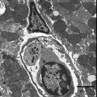
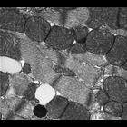
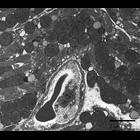
CIL:39756
NCBI Organism Classification
Mus musculus
Biological Process
none specified
Cellular Component
nucleus
Transmission electron micrograph of mouse cardiac muscle in the ventricle. A white blood cell is visible inside a blood vessel.
CIL:39765
NCBI Organism Classification
Mus musculus
Biological Process
Cellular Component
intercalated disc
Transmission electron micrograph of mouse atrial cardiac muscle showing a close up image of the intercalated discs, which support synchronized contraction of the cardiac muscle.
CIL:39754
NCBI Organism Classification
Mus musculus
Biological Process
transport
Cellular Component
none specified
Transmission electron micrograph of mouse cardiac muscle. Transport is visible in the ventricle across a space from one muscle cell to the adjacent muscle cell.
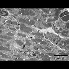
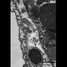
CIL:39781
NCBI Organism Classification
Mus musculus
Biological Process
muscle contraction
Cellular Component
I band
Transmission electron micrograph of ventricle tissue of a mouse cardiac muscle. This image shows the intercalated discs between the muscle fibers as well as many mitochondria and lipid droplets.
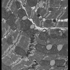
CIL:39727
NCBI Organism Classification
Mus musculus
Biological Process
regulation of cardiac muscle contraction
Cellular Component
sarcomere
Transmission electron micrograph of mouse cardiac muscle (ventricle). The image illustrates the myofibrils as well as a sarcomere in the area of fibrils between the two Z-lines. Z-lines link actin fi...
CIL:39775
NCBI Organism Classification
Mus musculus
Biological Process
cardiac muscle fiber organization
Cellular Component
mitochondrion
Transmission electron micrograph showing adjacent muscle fibers from the ventricle of a mouse cardiac muscle. This image shows the organization of muscle tissue, nucleus, cell walls and blood vessels...
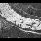
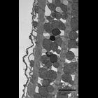
CIL:39760
NCBI Organism Classification
Mus musculus
Biological Process
atrial cardiac muscle tissue morphogenesis
Cellular Component
sarcoplasmic reticulum
Transmission electron micrograph of the periphery of an atria mouse cardiac muscle. This micrograph shows how rich cardiac tissue is with mitochondria reflecting its greater dependence on cellular re...
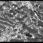
CIL:39757
NCBI Organism Classification
Mus musculus
Biological Process
ventricular cardiac myofibril development
Cellular Component
myofibril
Transmission electron micrograph of four longitudinally cut ventricle muscle fibers of mouse cardiac tissue. Muscle fibers are multinucleate celsl composed of myofibrils that contract when stimulated...
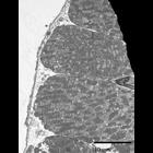
CIL:39764
NCBI Organism Classification
Mus musculus
Biological Process
atrial cardiac muscle tissue morphogenesis
Cellular Component
mitochondrion
Transmission electron micrograph of a blood vessel in the atria of a mouse cardiac tissue. This image is a close up of a red blood cell in a blood vessel.
CIL:39768
NCBI Organism Classification
Mus musculus
Biological Process
vesicle-mediated transport
Cellular Component
mitochondrion
Transmission electron micrograph of coated vesicles in mouse cardiac tissue. Coated vesicles have the materials bound to the receptor molecules incorporated within their membranes allowing the vesicl...
1
2
Next »
Results per page:
10
20
50
100