Alternate header for print version
Advanced search
Contributors
Help
Submit
Search
menu
Cell Process
Cell Component
Cell Type
Organism
Microbial
Alzheimer's
Data Sets
Center for Research in Biological Systems
University of California, San Diego
9500 Gilman Drive
La Jolla, CA 92093-0608, USA
Voice
: (858) 534-0276
Fax
: (858) 534-7497
Email
: dorloff@ncmir.ucsd.edu
Search Results for
Kathleen L Wilsen
(5 results)
(Not the results you were expecting?)
(Comments)
Still Images
Video/Animation
Z-Stack
Time Series
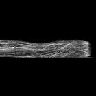
CIL:35287
NCBI Organism Classification
Lilium formosanum
Biological Process
pollen tube growth
Cellular Component
actin filament
Fixed Lilium pollen tubes were treated with fluorescent phalloidin to label filamentous actin, and confocal images obtained. A strongly labeled actin fringe near the growing tip is present. Other i...
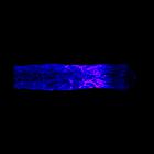
CIL:35290
NCBI Organism Classification
Lilium formosanum
Biological Process
pollen tube growth
Cellular Component
microtubule
Freeze substituted Lilium pollen tube immunolabeled for microtubules (blue>magenta) and imaged using laser scanning confocal microscopy. Shown is a projection of x-y slices revealing the distribution ...
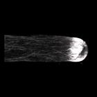
CIL:35285
NCBI Organism Classification
Lilium formosanum
Biological Process
pollen tube growth
Cellular Component
actin cytoskeleton
Freeze substituted Lilium pollen tube immunolabeled for filamentous actin and imaged using laser scanning confocal microscopy. Shown is a projection of z-slices revealing the dense sub-apical actin fr...
CIL:35286
NCBI Organism Classification
Lilium formosanum
Biological Process
pollen tube growth
Cellular Component
actin cytoskeleton
Freeze substituted Lilium pollen tube immunolabeled for filamentous actin and imaged using laser scanning confocal microscopy. Shown is a projection of x-z slices revealing the cortical distribution o...
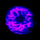
CIL:35291
NCBI Organism Classification
Lilium formosanum
Biological Process
pollen tube growth
Cellular Component
microtubule
Freeze substituted Lilium pollen tube immunolabeled for microtubules (blue>magenta) and imaged using laser scanning confocal microscopy. Shown is a projection of x-z slices revealing the cortical dist...