Alternate header for print version
Advanced search
Contributors
Help
Submit
Search
menu
Cell Process
Cell Component
Cell Type
Organism
Microbial
Alzheimer's
Data Sets
Center for Research in Biological Systems
University of California, San Diego
9500 Gilman Drive
La Jolla, CA 92093-0608, USA
Voice
: (858) 534-0276
Fax
: (858) 534-7497
Email
: dorloff@ncmir.ucsd.edu
Search Results for
Keith R. Porter
(9 results)
(Not the results you were expecting?)
(Comments)
Still Images
Video/Animation
Z-Stack
Time Series
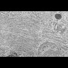
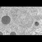
CIL:7722
NCBI Organism Classification
Microchiroptera
Biological Process
digestion
Cellular Component
rough endoplasmic reticulum
Stacks of rough endoplasmic reticulum (ER) cisternae seen in a transmission electron micrograph of a section through a secretory acinar cell from the bat exocrine pancreas. The rough ER is here involv...
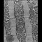
CIL:7567
NCBI Organism Classification
Myotis lucifugus
Biological Process
sarcomere organization
Cellular Component
mitochondrion
Cardiac muscle, particularly from the left ventricle, is rich in mitochondria since the heart requires an efficient continuous source of energy. In this highly magnified image, showing the length of t...
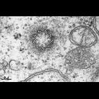
CIL:7726
NCBI Organism Classification
Gallus gallus gallus
Biological Process
centriole replication
Cellular Component
centriole
Cross-section of one of the centrioles of a diplosome and the surrounding region. The cells of a ciliated epithelium often produce many cilia, each of which grows from a 'basal body,' a structure that...
CIL:7725
NCBI Organism Classification
Rana pipiens
Biological Process
none specified
Cellular Component
cilium
CYT-165_3_Plate7Fig27_1200dpi.tif Electron micrograph of a thin section cut from the epithelium of the pharynx of frog, Rana pipiens. Structure of frog cilia in cross section J Richard McIntosh (Univ...
CIL:7739
NCBI Organism Classification
Chiroptera
Biological Process
cellular respiration
Cellular Component
mitochondrial crista
Here a mitochondrion is sliced approximately through its midplane, so both its outer membrane and the infoldings of its inner membrane are clearly seen. The organization of the inner membrane is parti...
CIL:7724
NCBI Organism Classification
Rattus
Biological Process
mitosis
Cellular Component
centriole
An electron micrograph showing that a centriole is constructed from a ring of nine triplet microtubules. Most animal cells contain two to four centrioles. In longitudinal view, a centriole looks like ...
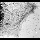
CIL:37242
NCBI Organism Classification
Gallus gallus
Biological Process
none specified
Cellular Component
endoplasmic reticulum
Classic TEM. First description of the endoplasmic reticulum. Whole mount of unfixed, dried chick embryo fibroblast. Embryonic chick cells were grown on a thin collodion film supported by a fine mesh ...
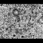
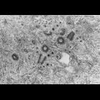
CIL:7723
NCBI Organism Classification
Cricetulus griseus
Biological Process
mitosis
Cellular Component
centriole
Four centrioles imaged in one electron micrograph of a thin section, cut from a Chinese hamster fibroblast grown in tissue culture. The two centrioles that appear circular are the 'mother' centrioles,...
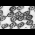
CIL:7751
NCBI Organism Classification
Cricetulus griseus
Biological Process
mitosis
Cellular Component
centriole
Centrioles multiply exactly once in preparation for cell division.In this remarkably rare image, four centrioles are seen in one electron micrograph of a thin section cut from a Chinese hamster fibrob...