Alternate header for print version
Advanced search
Contributors
Help
Submit
Search
menu
Cell Process
Cell Component
Cell Type
Organism
Microbial
Alzheimer's
Data Sets
Center for Research in Biological Systems
University of California, San Diego
9500 Gilman Drive
La Jolla, CA 92093-0608, USA
Voice
: (858) 534-0276
Fax
: (858) 534-7497
Email
: dorloff@ncmir.ucsd.edu
Search Results for
Susumu Ito
(14 results)
(Not the results you were expecting?)
(Comments)
Still Images
Video/Animation
Z-Stack
Time Series
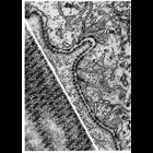
CIL:10946
NCBI Organism Classification
Siphonaptera
Biological Process
extracellular structure organization
Cellular Component
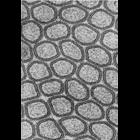
basal lamina
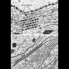
This electron micrograph shows the basal lamina along the midgut epithelium of the flea. Unlike the vertebrate basal lamina, which appears as a homogenous layer, the basal lamina in insects often app...
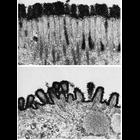
CIL:10937
NCBI Organism Classification
none specified
Biological Process
extracellular structure organization
Cellular Component
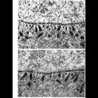
plasma membrane
These two micrographs show the outer cell surface of the epithelial cell border in intestine (upper) and stomach (lower) tissue. This surface, called the glycocalyx, is composed of negatively charged...
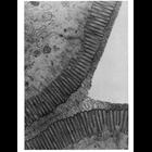
CIL:10929
NCBI Organism Classification
Felis catus
Biological Process
extracellular structure organization
Cellular Component
plasma membrane
This electron micrograph shows a region of the brush border of the cat intestine. Tufted and branched polysaccharide filaments, each a few nanometers thick, extend from the microvilli to make up the ...
CIL:10932
NCBI Organism Classification
Felis catus
Biological Process
extracellular structure organization
Cellular Component
plasma membrane
This electron micrograph highlights a darkly-stained glycocalyx rim of the brush border of the intestinal epithelium of the cat, stained en bloc with colloidal thorium. The glycocalyx is composed of ...
CIL:12063
NCBI Organism Classification
Felis catus
Biological Process
plasma membrane organization
Cellular Component
plasma membrane
This electron micrograph shows the plasma membrane of epithelial cells of the brush border of the cat intestine, obtained from a thin section parallel to the free surface. The ability to visualize ce...
CIL:11054
NCBI Organism Classification
Anura
Biological Process
nucleus organization
Cellular Component
nuclear envelope
Transmission electron micrographs of sections of tadpole oocyte show the potential for exchange between the nucleus and the cytoplasm at the nuclear envelope. Thin filaments that appear to traverse nu...
CIL:10987
NCBI Organism Classification
Unspecified
Biological Process
nucleus organization
Cellular Component
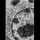
nucleus
Transmission electron micrograph of glutaraldehyde fixed pancreatic acinar cell showing the characteristic features of nuclear chromatin with this preparative method. Darkly staining heterochromatin (...
CIL:11405
NCBI Organism Classification
Myotis lucifugus
Biological Process
cellular respiration
Cellular Component
mitochondrion
Figures 224 (upper) and 225 (lower) from Chapter 7 (Mitochondria) of 'The Cell, 2nd Ed.' by Don W. Fawcett M.D. Mitochondrial granules range in size from 25 - 120 nm and vary by tissue and physiologi...
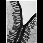
CIL:11201
NCBI Organism Classification
Siphonaptera
Biological Process
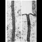
cell-cell junction organization
Cellular Component
septate junction
Epithelia of invertebrates contain zona continua and septate junctions not found in mammals. Left, zonula continua from gut epithelium of a flea; right, septate junction from the midgut epithelium of...
CIL:11832
NCBI Organism Classification
Arbacia
Biological Process
nucleus organization
Cellular Component
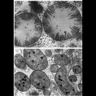
annulate lamellae
Transmission electron micrograph of stacked annulate lamellae from the oocyte of the sea urchin Arbacia. Annulate lamellae consist of sheets of membrane containing closely packed structures resemblin...
1
2
Next »
Results per page:
10
20
50
100