Alternate header for print version
Advanced search
Contributors
Help
Submit
Search
menu
Cell Process
Cell Component
Cell Type
Organism
Microbial
Alzheimer's
Data Sets
Center for Research in Biological Systems
University of California, San Diego
9500 Gilman Drive
La Jolla, CA 92093-0608, USA
Voice
: (858) 534-0276
Fax
: (858) 534-7497
Email
: dorloff@ncmir.ucsd.edu
Search Results for
Tatyana Svitkina
(108 results)
(Not the results you were expecting?)
(Comments)
Still Images
Video/Animation
Z-Stack
Time Series
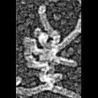
CIL:24804
NCBI Organism Classification
Xenopus laevis
Biological Process
branching of actin filaments
Cellular Component
actin cytoskeleton
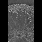
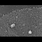
Differential response of lamellipodial actin network to latrunculin a (LA), a binder of monomeric actin. This electron micrograph of a Xenopus keratocyte)lamellipodium treated with LA (0.1 μM for 30...
CIL:24805
NCBI Organism Classification
Xenopus laevis
Biological Process
branching of actin filaments
Cellular Component
actin cytoskeleton
Differential response of lamellipodial actin network to latrunculin a (LA). Electron micrograph of a Xenopus fibroblast lamellipodium treated with LA (0.25 μM for 10 min) reveals actin depletion fro...
CIL:24810
NCBI Organism Classification
Xenopus laevis
Biological Process
latrunculin a treatment
Cellular Component
lamellipodium
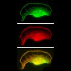
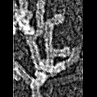
Localization of XAC XAC (Xenopus ADF/cofilin) to posterior regions of depolymerization-resistant actin brush. Fluorescence microscopy of lamellipodia of Xenopus keratocytes after latrunculin a (a bin...
CIL:24784
NCBI Organism Classification
Xenopus laevis
Biological Process
actin filament organization
Cellular Component
lamellipodium
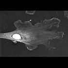
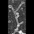
Localization of XAC (Xenopus ADF/cofilin) in Xenopus fibroblasts. Immuno-EM with XAC antibody at low magnification. High magnification view is available at CIL 24785. Nucleus and surrounding regions...
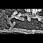
CIL:24788
NCBI Organism Classification
none specified
Biological Process
branching of actin filaments
Cellular Component
lamellipodium
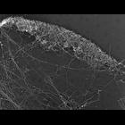
Multiple branching of actin filaments in lamellipodia of vertebrate fibroblasts. This image shows an overview of the leading edge, and CIL 24789 shows enlargements of local regions of this platinum re...
CIL:35064
NCBI Organism Classification
none specified
Biological Process
branching of actin filaments
Cellular Component
actin cytoskeleton
Electron micrograph of keratocyte or fibroblast lamellipodial actin network after unprotected extraction. All examples demonstrate frequent branching of actin filaments. Image corresponds to a singl...
CIL:35067
NCBI Organism Classification
none specified
Biological Process
branching of actin filaments
Cellular Component
actin cytoskeleton
Electron micrograph of keratocyte or fibroblast lamellipodial actin network after unprotected extraction. All examples demonstrate frequent branching of actin filaments. Image corresponds to a singl...
CIL:35068
NCBI Organism Classification
none specified
Biological Process
branching of actin filaments
Cellular Component
actin cytoskeleton
Electron micrograph of keratocyte or fibroblast lamellipodial actin network after unprotected extraction. All examples demonstrate frequent branching of actin filaments. Image corresponds to a singl...
CIL:35061
NCBI Organism Classification
none specified
Biological Process
branching of actin filaments
Cellular Component
actin cytoskeleton
Electron micrograph of keratocyte or fibroblast lamellipodial actin network after unprotected extraction. All examples demonstrate frequent branching of actin filaments. Image corresponds to a singl...
CIL:34899
NCBI Organism Classification
none specified
Biological Process
branching of actin filaments
Cellular Component
actin cytoskeleton
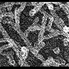
Improved visualization of actin filament branching in lamellipodia. EM of keratocyte or fibroblast lamellipodial actin network after cytochalasin D treatment (0.2 μM for 30 min or 0.5 μM for 10 min...
1
2
3
4
5
6
7
8
9
...
11
Next »
Results per page:
10
20
50
100