Alternate header for print version
Advanced search
Contributors
Help
Submit
Search
menu
Cell Process
Cell Component
Cell Type
Organism
Microbial
Alzheimer's
Data Sets
Center for Research in Biological Systems
University of California, San Diego
9500 Gilman Drive
La Jolla, CA 92093-0608, USA
Voice
: (858) 534-0276
Fax
: (858) 534-7497
Email
: dorloff@ncmir.ucsd.edu
Search Results for
Vincent Pasque
(6 results)
(Not the results you were expecting?)
(Comments)
Still Images
Video/Animation
Z-Stack
Time Series
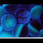
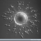
CIL:39469
NCBI Organism Classification
Xenopus laevis
Biological Process
oocyte construction
Cellular Component
nucleus
Fluorescent micrograph of stage V-VI Xenopus laevis oocytes surrounded by thousands of follicle cells, as visualized by Hoechst staining.
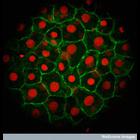
CIL:39470
NCBI Organism Classification
Danio rerio
Biological Process
none specified
Cellular Component
nucleus
Confocal projection of Zebrafish yolk cells expressing H2B-cherry (red) to mark the nucleus and a GFP membrane marker (green) to show the outline of the cells.
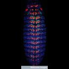
CIL:39468
NCBI Organism Classification
Drosophila
Biological Process
gene expression
Cellular Component
Pax 3/7 gene
Confocal micrograph of a Drosophila embryo stained for Pax3/7 (cyan), eve, horse radish peroxidase (red) and counterstained with DAPI (blue). Pax3/7 genes encode a class of transcription factors that...
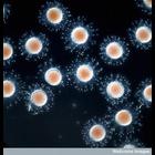
CIL:39466
NCBI Organism Classification
Ascidiacea
Biological Process
embryo development
Cellular Component
cell surface
Early ascidian (sea squirt) embryos visualized by differential interface contrast (DIC) microscopy. Ascidians are used as a model for developmental research. Their simple embryonic development is rapi...
CIL:39471
NCBI Organism Classification
Ascidiacea
Biological Process
embryo development
Cellular Component
cell surface
Early ascidian (sea squirt) embryos visualized by differential interface contrast (DIC) microscopy. Ascidians are used as a model for developmental research. Their simple embryonic development is rapi...
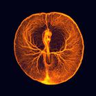
CIL:41734
NCBI Organism Classification
Gallus gallus
Biological Process
vasculogenesis
Cellular Component
none specified
This image of the vascular system of a chicken embryo taken two days after fertilization is a composite of two different images taken with an upright fluorescent dissecting scope. The egg's shell was...